Anatomy Of Ribs Posterior - Fusion Of The Posterior Aspect Of The 3rd And 4th Right Ribs Download Scientific Diagram - Review the anatomical characteristics of the rib and ribcage in this interactive tutorial and test your knowledge in the quiz.
byAdmin-
0
Anatomy Of Ribs Posterior - Fusion Of The Posterior Aspect Of The 3rd And 4th Right Ribs Download Scientific Diagram - Review the anatomical characteristics of the rib and ribcage in this interactive tutorial and test your knowledge in the quiz.. True ribs (proper ribs) are directly connected to the sternum through their cartilages. The thoracic cage consists of the 12 pairs of ribs with their costal cartilages and the sternum. In vertebrate anatomy, ribs (latin: Head of rib articulates with vertebra ribs move as a unit to accommodate breathing intercostal spaces = (spaces between ribs) • • •. This muscle is present posteriorly within the thoracic wall.
The thoracic vertebrae are located in the thorax posterior and medial to the ribs. Each rib articulates posteriorly with two thoracic vertebrae by the costovertebral joint. Common characteristics of the ribs figs. Includes images, video, and free quiz. Illustrations in anterior and posterior view of male torso and back, allowing the lines and regions used in surface anatomy to be displayed (midclavicular line, midline, pectoral region, sternal region.) ribs:
The Anatomy Of The Ribs And The Sternum And Their Relationship To Chest Wall Structure And Function Sciencedirect from ars.els-cdn.com Exposure of the posterior mediastinum is through the bed of the seventh or eighth ribs. Skeletal system anatomy and physiology nurseslabs. The subclavian artery and brachial plexus cross the rib posterior to anterior scalene muscle attachment and then run in contact with the bone on their way to the upper limb. Continue scrolling to read more below. The rib below that is rib 2, and it connects to the t2 thoracic vertebra, and so on. Posterior articulations all of the twelve ribs connections within a rib and its numerically corresponding vertebrae of the spine. Includes images, video, and free quiz. Anatomical name for floating ribs, anatomical term ribs, anatomical word for ribs, anatomy ribs quiz, ribs anatomical position, human anatomy anatomy of shoulder 12 photos of the anatomy of shoulder anatomy of nerves in shoulder, anatomy of posterior shoulder dislocation, anatomy.
This incision may be continued across the costal margin to open the abdominal cavity as in.
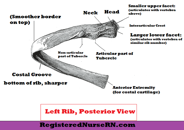
Skeletal system anatomy and physiology nurseslabs. Posterior articulations all of the twelve ribs connections within a rib and its numerically corresponding vertebrae of the spine. Head of rib articulates with vertebra ribs move as a unit to accommodate breathing intercostal spaces = (spaces between ribs) • • •. Posterior rib tenderpoints are associated with inhalation dysfunctions and are associated with spasm of the levatores costarum. The part of the muscle is thought to depress the ribs. They form the region of the spinal column inferior to the cervical vertebrae of the neck and superior to the lumbar vertebrae of the lower back. However, they do not attach directly to the sternum anteriorly, and instead, attach to the. The posterior end is composed of head, neck, and tubercle. Each rib articulates posteriorly with two thoracic vertebrae by the costovertebral joint. 1.3 ribs anatomy and somatic dysfunctions. The ribs stretches posteriorly from thoracic vertebrae to the anterior lateral edges of the sternum. Major landmarks of a typical rib are the following: Illustrations in anterior and posterior view of male torso and back, allowing the lines and regions used in surface anatomy to be displayed (midclavicular line, midline, pectoral region, sternal region.) ribs:
In the anatomical position, the scapula overlies the second to seventh ribs on the posterolateral aspect of the chest wall. Serratus posterior superior and inferior. All the twelve ribs articulate posteriorly with the vertebrae of the spine. The thoracic vertebrae are located in the thorax posterior and medial to the ribs. This incision may be continued across the costal margin to open the abdominal cavity as in.
Ribs Pictures Anatomy Anatomy Body Maps from post.healthline.com The thoracic vertebrae are located in the thorax posterior and medial to the ribs. Illustrations in anterior and posterior view of male torso and back, allowing the lines and regions used in surface anatomy to be displayed (midclavicular line, midline, pectoral region, sternal region.) ribs: Costae) are the long curved bones which form the rib cage, part of the axial skeleton. Each rib articulates posteriorly with two thoracic vertebrae by the costovertebral joint. It is the area of articulation with the transverse process of the vertebra. Roughly speaking, this is the area of the chest. Ten of the twelve ribs connect to strips of hyaline cartilage on the anterior side of the body. Medical illustrations muscle, vascular, abdominal wall.
Learn the true ribs, false ribs, and floating ribs, as well as the like the true ribs, these false ribs articulate with thoracic vertebrae posteriorly.
Represents the anatomy of the ribs and muscle attachments. Skeletal system anatomy and physiology nurseslabs. Ribs 3 to 9 are considered typical ribs. The thoracic vertebrae are located in the thorax posterior and medial to the ribs. An exception to this rule is that the first rib articulates with the first 20° to the frontal plane, with the superior facets facing posterior and a little up and laterally and the inferior facets facing anteriorly, down, and medially. It is split into ibrahim, af and darwish: The posterior abdominal wall is a musculoskeletal structure formed by the posterior abdominal muscles posteriorly by the lumbar vertebrae, muscles, and fascia. In this video, you will learn the bony features of typical and atypical ribs. Common characteristics of the ribs figs. Each rib articulates posteriorly with two thoracic vertebrae by the costovertebral joint. Ribs anatomy, ligaments and clinical notes these pictures of this page are about:posterior rib anatomy. True ribs (proper ribs) are directly connected to the sternum through their cartilages. Further details of its anatomical relations and muscle attachments can be found in its own section in this text.
Continue scrolling to read more below. Vertebral column, ribs, and the sternum. Medical illustrations muscle, vascular, abdominal wall. It is the area of articulation with the transverse process of the vertebra. Head, neck, tubercle, and body of a rib.
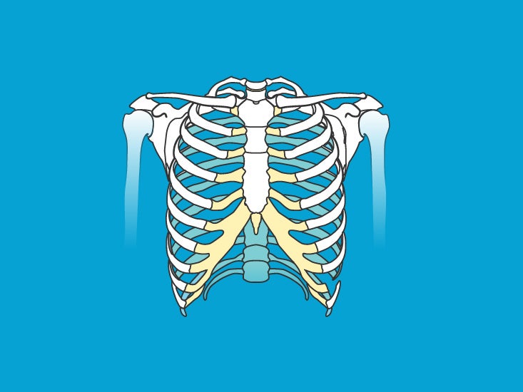
Ribs Anatomy True Ribs False Ribs Floating Ribs Typical And Atypical Ribs from www.registerednursern.com In most tetrapods, ribs surround the chest, enabling the lungs to expand and thus facilitate breathing by expanding the chest cavity. The part of the muscle is thought to depress the ribs. Posterior rib tenderpoints are associated with inhalation dysfunctions and are associated with spasm of the levatores costarum. The ribs are a set of twelve paired bones which form the protective 'cage' of the thorax. The posterior abdominal wall is a musculoskeletal structure formed by the posterior abdominal muscles posteriorly by the lumbar vertebrae, muscles, and fascia. However, they do not attach directly to the sternum anteriorly, and instead, attach to the. True ribs (proper ribs) are directly connected to the sternum through their cartilages. Roughly speaking, this is the area of the chest.
Exposure of the posterior mediastinum is through the bed of the seventh or eighth ribs.
The ribs are elastic arches of bone, which form a large part of the thoracic skeleton. Exposure of the posterior mediastinum is through the bed of the seventh or eighth ribs. Anatomical name for floating ribs, anatomical term ribs, anatomical word for ribs, anatomy ribs quiz, ribs anatomical position, human anatomy anatomy of shoulder 12 photos of the anatomy of shoulder anatomy of nerves in shoulder, anatomy of posterior shoulder dislocation, anatomy. It is split into ibrahim, af and darwish: In the anatomical position, the scapula overlies the second to seventh ribs on the posterolateral aspect of the chest wall. In most tetrapods, ribs surround the chest, enabling the lungs to expand and thus facilitate breathing by expanding the chest cavity. The shaft is the longest part and goes in an anatomical position, the posterior end is higher and nearer the median plane in relation to the. Skeletal system anatomy and physiology nurseslabs. All 12 pairs of ribs attach to the building blocks of the spine (vertebrae) in the back. Posterior left rib fractures with injuries and nonunion of. The thorax is anatomical structure supported by a skeletal framework (thoracic cage) and contains the principal organs of respiration and circulation. The ribs form the main structure of the thoracic cage protecting the thoracic organs, however their main function is to aid respiration3. Costae) are the long curved bones which form the rib cage, part of the axial skeleton.
The ribs are elastic arches of bone, which form a large part of the thoracic skeleton anatomy of ribs. Exposure of the posterior mediastinum is through the bed of the seventh or eighth ribs.